

Andrej Stancak
-
Posts
1,262 -
Joined
-
Last visited
Content Type
Profiles
Forums
Events
Store
Posts posted by Andrej Stancak
-
-
Mr. Frazier will never tell the full truth about the events he witnessed during the assassination Friday. I read his autobiography book and watched or listened to perhaps all his interviews he made over the long period of time since that fateful Friday, November 22, 1963.
Mr. Frazier still experiences symptoms of postraumatic stress disorder which he seems to have developed based on the hardship he sustained as a child, especially the abuse from his stepfarther, and triggered by his situation on that Friday. The symptoms of PTSD include lapses in memory, often very selective and related to the stressful event, avoiding conversations on the topic, and emotional distress experienced when recalling the events. Mr. Frazier has manifested all of these symtoms. The life threatening event was the interrogation at the Dallas police headquarters which threatened to make him an accomplice to the assassination by his assocation with Lee Oswald.
Mr. Frazier may genuinly not remember that he had briefly seen Lee in the doorway even if Lee was there, less than 3 feet away from him.
-
There cannot be any doubts about the authenticity of Hosty's statement regarding Oswald going out to watch the "P parade". The narrative containing this statement was written on a sheet of paper used by the Dallas Police to take affidavits from witnesses. Hosty in his book "Assignment Oswald" confirmed that he grabbed such a sheet from Fritz's desk at the start of the interrogation session at 3.15PM, November 22. Hosty notes on Oswald's whereabouts, discovered by Bart Kamp, match Hosty's own book exactly except the small detail about going out to watch the "P. parade". Briefly, the notes and the book say that Oswald was eating his lunch in the first floor lunchroom during the time when the motorcade passed the building.
It is possible that Lee Oswald heard the excitement and the crowd noise when the motorcade approached Dealey Plaza and began turning onto Houston street.
I made a time reconstruction of Lee's trajectory starting in Domino room and ending in the doorway which can be found here (my post dated August 29, 2020):
https://educationforum.ipbhost.com/topic/22247-prayer-man-is-a-man/page/14/
This time reconstruction allows Lee to leave Domino room within a 9-second window starting 3 seconds after the motorcade turned onto Houston street and still be filmed by Wiegman as standing in the doorway. Thus, it appears that Lee walked out to watch "P. parade" but he came in to the doorway only around the time of the third shot or even a moment later.
-
There may have been multiple line-ups in which Lee Oswald participated. The principle of the line-ups was always the same - Lee had to look odd in a line-up.
In one of the line-ups, Lee was paraded in a T-shirt with teenagers around him in regular shirts, and Lee complained:
https://www.youtube.com/watch?v=20SzdIBn51Q
In another line-up, older men possibly dressed in suits were presented along with Lee Oswald:
https://www.maryferrell.org/photos.html?set=WCD-LINEUPS
I was not too glad seeing one of the Forum members criticising Joseph's presentation and, by extension, his work. "Into the nightmare" is one of the most revealing and best researched books in the realm of JFK's assassination. I read it twice already, and will read it again to appreciate all details and valuable bits of information one cannot find anywhere else, e.g., the account of Joseph's interview with Tippit's father.
-
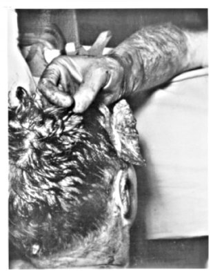
James Jenkin's index finger points directly to the area where he saw the open head wound before the scalp was retracted. The wound as he remembers it was sized 3x2 inches. The wound would be largely in the occipital bone and spread somewhat to the parietal bone.
The image below is a screenshot of a frame at 29:45 in video interview with James Jenkins, assistant pathologist at the medical school of the Bethesda Naval Hospital.

-
10 hours ago, Pat Speer said:
Oh my. If you really think the back of the head is flat, I suggest you take this tool called a hand and place it on the back of your head. You will quickly realize that the area stretching from the suture on down 2 1/2 inches is far from flat.
Human heads differ in shapes and the occipital-parietal region is one of the examples. Some people have rounded occipital bones, other people have the back of the head straight and tall. This is why I showed the view of President Kennedy in which the back of his head is clearly visible.
Unless your head model matched Kennedy's head in size and shape - particularly in the back portion of the head, you would not be able to conclude whether the bone was flat enough or large enough to give Harper fragment. It cannot be decided without testing it. I doubt that touching own head would help too much in this process; this method looks too simple to me, given the differences in size and shape of human heads.
-
As to what head wound the Parkland doctors and nurses could see on Kennedy's head, Dr Aguilar's narrative explains the situation in Trauma Room One. The head wound could not be seen from a side view and therefore, it could not be located on the convexity of the parietal bone, and it took some effort to observe the head wound in the back of the head:
"Author David Lifton reported that Parkland emergency nurse Audrey Bell claimed that JFK's skull wound "was so localised at the rear of JFK's skull that, from her position on the right side on the right hand side, with Kenned's lying face up, she couldn't see any damage". It is certainly likely that a blown-out skull wound on the right side would have been visible to witnesses standing on that side. But had the skull defect been more on the back of JFK's head, rather than on the side - which, as we'll see is what virtually all the witnesses first reported - then some sense can be made of Jenkins' and Bell's comments that the wound was not visible to side witnesses. It would also help explain the similar, previously suppressed, report from a witness who was present at JFK's autopsy - General Phillip C. Wehle, Commanding Officer of the military District of Washington, D.C. After interviewing Wehle in 1978, House Select Committee on Assassination (HSCA), councel D. Andy Purdy J.D., reported that, (Wehle) noted the wound was in the back of the head so he would not see it because the President was lying face up..." (page 179)
"The Boston Globe reported that "some (Parkland) doctors doubted the extent to which a wound to the rear of the head would have been visible since the President was lying supine with the back of his head on a hospital cart."
The Globe immediately refuted that speculation, reporting. "But others, like (Dr. Richard Dulaney) and (neurosergeon Dr. Robert) Grossman, said the head at some point was lifted up, therefore exposing the head wound". Similarly, author David Lifton reported that Parkland emergency nurse Audrey Bell, who couldn't see JFK's head wound though she was standing on the right side, asked Dr. Perry. "Where was the wound?", Perry pointed to the back of the President's head and moved the head slightly in order to show her the wound". During sworn interviews with the ARRB in 1998, Dr. Paul Peters reported , "(anesthesiologist Dr. Marion T.) Jenkins said, "Boys, before you think about opening the chest, you'd better step up here and look at his brain." And so at that point I did step around Dr. Baxter and looked into the President's head...". The ARRB's Gunn inteviewed neurosurgeon Robert Grossman, M.D. on March 21, 1997. reporting, "He (Grossman) and Kemp Clark (Chairman of Neurosurgery at Parkland) (sic) together lifted President Kennedy's head so as to be able to observe the damage to the President's head." (page 193).
Quoted from Aguilar, G, The converging medical case for conspiracy, In: Murder in Dealey Plaza, J. Fetzer (ed.), Chicago Press, 2000.
-
Pat:
I read Riley's report. His report is not really a proof of anything. His claim that occipital bone is devoid of vascular grooves is false (Dr Mantik has discussed it in detail in his book) and I myself have checked quite many plates in current anatomy books, and of course, vascular grooves can be found on the interior of the squama of occipital bone.
It is quite a bit of serious work to model and fit Harper fragment into a head model, and while I started some preparatory work, this project is far from completion, and I therefore cannot show the location I currently think is the correct one because this location may change in the later stages of the project, and I do not wish to spread information about my unfinished project. However, even my preparatory reading was sufficient to be able to see obvious mistakes in the assumed parietal location of Harper fragment. The most obvious mistake is that the parietal bone is convex, angulated, while Harper fragment is a flat piece of bone. Therefore, it would not fit the parietal bone.
It is encouraging that we both can agree on the fact that Harper fragment was a piece of JFK's skull and that it was blasted out from his head in such a way that a hole would have been seen on JFK's head. If it would have been on the parietal bone, all Parkland staff would see it at once and would locate it by pointing to the side of their head between vertex and the ear. But they did not do that. The hole was elsewhere - in the back of the head, this is where they pointed to.
Here is a posterior view of human skull giving a good indication of how convex the parietal bone is, unlike the squama of the occipital bone above the superior nucha which region of the skull is flat. Harper fragment would only fit a flat bone.

This photograph (Kennedy meeting miners in West Virginia, 1960) nicely shows the (flat) back of Kennedy's head.

-
5 minutes ago, Pat Speer said:
In any event I tackled this myself and Riley was obviously correct--there's no way the Harper fragment is occipital bone.
There are obviously conflicting views between the professional pathologists who were able to inspect the bone fragment itself rather than to work out the locations from the photographs, Jack C. Harper, Gerard Noteboom, and A.B Cairns (chief of pathology department at Methodist Hospital, Dallas), and the experts called by the HSCA. Dr Mantik did a good job to dispute the views of J.J Riley and J. Angel who likewise were experts. My point is that none of the reports by the HSCA experts or Dr Mantik are based on a realistic reconstruction of the fragment and its fitting into a model of JFK's head, and none of their work is convincing enough. It is not possible just to claim something, it is necessary to prove it. The HSCA did all to obfuscate and not really investigate the medical evidence (e.g., forcing Dr Humes to shift the entry wound from the level of external occipital protuberance about 10 cm superior to the parietal bone), so Riley's and Angel's views are irrelevant today unless they presented convincing proofs of the parietal location which they did not.
-
5 hours ago, Sean Coleman said:
note Harper fragment included in this imaginification of a diagram
Sean:
Harper fragment is a flat piece of bone with only its "beacon" being curved sligthly inward. This fragment could not originate from the parietal bone as shown in the sketch in your post where the convexity of the skull is too much pronounced. The only region of the skull where this fragment could come from was the occipital bone above the occipital protuberance which bone was remarkably flat on Kennedy's skull. The curved "beacon" is from the lateral portion of the upper part of squama of occipital bone as it curves anteriorly, basically it is a part of the wall of the transverse sulcus. The two lines on the interior surface of Harper fragment are sulci for, probably, the blood vessels. While Dr Mantik and the Dallas doctors who photographed Harper fragment were correct in placing the fragment into occipital bone, Dr Mantik's fit of Harper fragment was not correct, in my opinion.
I have started a 3D model of Harper fragment and downloaded a large number of CT head scans (512 heads) to find the head volume matching Kennedy's head the best, to test the goodness of fit of the realistic 3D model of Harper fragment in various hypothesised locations. However, the parietal bone location of Harper fragment would be an unlikely solution owing to the convexity of the parietal bone. From what I read about Harper fragment, no one really accomplished a proper analysis to fit this fragment into Kennedy's skull, possibly due to limited technology at that time.
-
Eddy:
Doctor Humes in his ARRB testimony admitted that several centimeters of scalp were missing in such a way that the scalp could not be closed. As Parkland doctors did not see any head wound on the lateral convexity of the head, it could have been only the back of the head showing a deficit in scalp tissue. It is true that Dr Humes did not confer in his ARRB deposition which part of the head lacked the scalp, he simply dodged the question.
-
10 hours ago, Jonathan Cohen said:
This is the same reason Tom Wilson's claims of being able to detect widespread alteration in the assassination photo record never rang true to me -- he was studying multiple-generation copies, and not the original evidence.
Jonathan:
I am sure you are wrong here. I have a part of Tom Wilson's archive (sent to me by Don Philips) as I have been engaged in the reconstruction of his image analysis method. Tom Wilson had Mary Moorman's photograph made from the original negative of the polaroid picture. The copy of e.g., MM photograph I have is a high-resolution copy of Tom's photograph. The autopsy photographs Tom had analysed were high-quality pictures he received from Cyril Wecht. Some of the photgraphs (e.g., those showing Dealey Plaza) were given to Tom by Oliver Stone when Oliver briefly hired Tom's services in 1991 during shooting of his JFK film.
Tom Wilson was very well aware of the importance of having the best quality original picture for any photographic analysis.
The autopsy photgraphs available at jfkassassination.org are certainly not copies made from a book. Copies made from book would either show a grid-like raster if scanned at high resolution, or would appear too smooth if a filter would be applied to get rid of the raster. The photograph with apparently intact hair in the back of the head (the first photograph shown in this thread) even shows thin line scratches around the surgeon's arm which proves that details were maintained; these autopsy pictures were made from a good quality copies of autopsy photographs. Robin Unger could advise as to the origin of the autopsy pictures displayed on jfkassassinationgallery.org.
Late edit: I should add that the brightness-coding analysis method used in this thread is the same which Tom Wilson had used. However, Tom was able to program it all by himself while I use a freely available program ImageJ that was developed for analysis of medical and biological images. I have invested a lot of time, funds and effort into the hardware part of Tom Wilson's method (shooting both in infra-red and visible spectrum of light using a sensitive industrial camera, illuminating the enlargements of images with strobe light at different frequencies, enlarging the brightess coding to 12 bits instead of 8 bits), and currently I cannot say what is the added value of each of these elements. This is a very time consuming project and I am afraid it will need to wait until my not-so-distant retirement to be able to get to the bottom of Tom's method.

-
9 hours ago, Eddy Bainbridge said:
Hi Andrej, what do you have against the simpler explanation that the BOH photo is after the reconstruction? If you read Hulmes reaction to the photo (HSCA) it pretty much confirms he had requested a photo to show the (small) rear headwound, but it didn't. It is my view that it didn't because the scalp sagged over the hole. The large blowout in the rear had been sealed with a piece of rubber and the scalp sewn up. I think its possible that what you are picking up is stretched scalp , where some follicles are further apart than before the injuries.
Eddy:
there was no scalp in the open wound in the back of the head. There was no scalp to be stretched over that open wound through which the brain matter and fluids were discharded after the fatal head shot.
Humes could not see the opening in the back of the head not because of scalp retraction but owing this picture being blackened in the back of the head. If there would be retracted scalp in the back of the head, we would still see the texture of hair.
Your note indicates that you think the timing of the photograph was after the morticians completed embalming the body (sealing the wound with rubber...). However, I see physicians still taking measurements of the back, meaning it is an autopsy photograph.
-
10 hours ago, Pat Speer said:
They didn't study the photos posted on this thread. They had the negatives and the original prints to work with. The photos you are looking at are probably photos of photos that had been printed in books. So let's see. There's the original negative. Then a photo made from that negative. Then a photo of that photo that was printed in a book. Then a digital copy scanned from that book, or perhaps even a digital copy scanned from a photo of a photo in that book. Either way, that's a long way down, a long way in which the relative contrast within the photo could have been changed, and details could have been lost.
Pat:
The most recently shown photograph (posted on Sunday, June 19) actually shows very good details over the entire picture excluding the back of the head, and including e.g., the details of hair in the upper part of the scalp. President's back and and physician's hands are sharp too. The only part of the photograph showing only flat black colour with no depth is the right and left back of the head down to the neck. If degradation of the original photograph due to overcopying would cause dark patches, the degradation of the same strength would be seen uniformly across the picture. However, it is not. There is simply no signal in the back of the head. However, it is not only the lack of details but also the black colour of the patch. It is not possible to suddenly have a dark patch in one region of the head (alluding to no external or ambient light present) but not in the neck or on the top of the head.
I would love to be able to view the certified copies of autopsy images, however, this is unlikely to happen. Therefore, until someone shows this picture with all details in the back of the head (including Hume's entry wound to the right of external occipital protuberance) this particular image remains a crude alteration of the autopsy evidence and a proof of a coverup.
-
I do not understand why the HSCA did not point to the blatant tampering with the autopsy picture shown in my last post. It would be more difficult to show the presence of a hole in the back of the head in the picture showing hair in that area (the first autopsy picture in this thread), however, the image manipulation in the latter picture is far too obvious. The commissioners should have pointed to the forgery and ask for explanations; it would still be possible, back in 1978, to trace the history of this forgery right to the perpetrators.
The alterations made in both autopsy pictures discussed in this threat invalidates the results of the autopsy in its entirety. Moreover, the fact that the manipulations in these pictures occurred in the back of the head prove that the back of the head was the critical head region that the perpetrators of this crime wished to cover up.
-
The photograph I consider as being crudely retouched is the one below (downloaded from jfkassassinationgallery.org). Please note a non-transparent black patch over both the left and right back of the head. The left panel shows the original photograph, and the right panel shows the same photograph after light has been added to dark tones. There is no natural explanation for the occurrence of this dark patch.


-
45 minutes ago, Pat Speer said:
Oh my. Robert Knudsen was not an autopsy photographer and did not take photos of Kennedy's autopsy. Heck, he wasn't even at the autopsy. The autopsy photographer was John Stringer, and his assistant, who took a few photos that were overexposed by the SS, was Floyd Riebe.
Maybe I misunderstood, however, this is what I got from JFK Absolute Proof by Robert Groden:
"Pressed into service as an autopsy photographer on the night of November 22, 1963, White House photographer Robert Knudsen was questioned by the House Select Committee on Assassinations in 1978." (p.164).
I interpret this sentence as Knudsen being present at the autopsy and taking pictures on behalf of the White House, in parallel to the official photograhers Stringer and Riebe.
-
37 minutes ago, Michael Crane said:
Hello Andrej,
It's my belief that the reason the photographic panel did not find any forgeries in the autopsy photographs was because the manipulators took a picture of the picture you provided.This would result in not being able to identify any forgeries.
Michael:
I agree completely. The picture, in my view, was manipulated by adding a texture alluding to hair, and the picture was re-shot while selectively underexposing the right side of the head during the positive process ( I used to employ such simple masking myself in my young years if I wanted to brighten or darken selected parts of the photograph). The right portion of the scalp looks unexplicably dark compared to the part of the scalp over the vertex and to the left of vertex.
The autopsy photograph analysed here is labelled "This is the photograph that Robert Knudsen confirmed as fake", on page 166 in JFK Absolute truth by Robert Groden. Robert Knudsen was the White House photographer tasked with taking photographs during the autopsy.
In other photographs, chief autopsy photographer John Stringer conferred to the ARRB under oath that the film containing autopsy pictures shown to him was not the type of film he had used in 1963. That would mean that the film shown to him was photographed by shooting forged pictures.
However, we are fortunate to have at least this type of fake. The worse fakes are those having portions of the back of the head completely retouched. I can try to analyse those too, however, I do not feel this could yield anything.
-
There are numerous threads on medical evidence and autopsy data on this forum, and as I do not know which thread to choose, I decided to start a new one.
The lack of a wound on the back of President's head in official autopsy photographs is in stark contrast with numerous testimonies by Parkland medical staff who were consistent in observing such wound while President was attended to in trauma room 1. While some autopsy photographs have the back of the head crudely retouched, there may be one or couple pictures which could still contain original information pertaining to the wound in the back of President's head.
The picture below appers to be just such picture (downloaded from jfkassassinationgallery.org) (left):

I have added a bit of light to the dark tones which resulted in the image in the right panel.
The logic of the problem suggests that in case of intentional alteration of this autopsy picture to hide the occipital-parietal wound, a texture in form of human hair would be added to mask the dark space represented by the wound. If comparatively light-coloured lines have been added to otherwise very dark area (hollow space of the wound), we should see a different distribution of very dark tones over the area of the wound compared to the area of intact hair. Basically, the hair fibres over the tampered area would not have have the correct vertical depth and would end abruptly in the dark void of the head wound.
I have uploaded this image to ImageJ program which allows visualisation of the brightness of an image as a depth, creating a pseudo 3D image in which light tones are on the surface of the 3D structure and the dark tones in the depth of the 3D structure.
Here are the black-and-white and coloured representations of the image above (the enhanced one):


As brightness adds another dimension to the image, the photograph becomes a 3D object and can therefore be tilted, rotated etc. In the coloured image, please note that the very dark area next to the surgeon's thumb is represented as being located deep and it is painted with dark blue or black colour. This area of the head is likely devoid of any tissues, a cavity.
It is possible to view the histogram of brightness across the height of the image which allows to view the configurations of the very dark pixels across the picture; those would likely be in areas devoid of any tissues.

In the histogram image above, the top of the patch would be the most superficial layer of comparatively bright pixels, and the bottom of the patch would represent the distribution of comparatively dark pixels. The photograph is viewed as if from the side with the left corner being at President's neck and the right corner ending at the surgeon's elbow. Please note the very dark area corresponding to the region of the head next to the surgeon's finger. However, the occipital area of the head down to a line at approximately the level of external occipital protuberance is also very dark.

The picture above shows the map of dark pixels in the right parieto-occipital region of the head. Basically, we see the bottom of 3D brightness-coded photograph. Please note that hair above the neck shows intermediate level of blue-violet colour (skin), while the region above of what may be the hollow area has clearly a darker "bottom" and connects to the dark area next to the surgeon's finger. In my humble opinion, the black-coded region in the photograph is related to a void owing to the missing tissue in the occipital-parietal region of the head. Interestingly, there appears to be a very bright spot interrupting the lower contour of the dark area. Of course, this spot would be bright when viewed from the top and it would be hard to pinpoint it as the neighboring spots would also be of comparatively light colour. However, unlike other light-coloured lines on the bright surface of the photograph, this spot has no dark bottom, meaning it is bright over the entire depth. One explanation would be that it is a metal object lodged in the lower aspect of the head wound.

The "bottom" of the brightness coded pseudo-3D picture was overlaid onto the original photograph. This visualisation may actually point to the President's occipital-parietal wound.
Notably, the texture of normal hair in the lower back region of the head differs from the texture of hair in the area overlaying the purported hollow space.

The hair fibres in the lower part of the scalp ("normal hair texture") create vertical columns with a continuous gradation of brightness, and have a shape of Eiffel tower. In contrast, the columns of hair in the area overlaing the void have only a comparatively bright top and fall sharply to the very dark levels with almost no gradations of brightness (a skyscraper). This would be consistent with painting comparatively bright patches over a very dark region of the photograph overlaying a cavity.
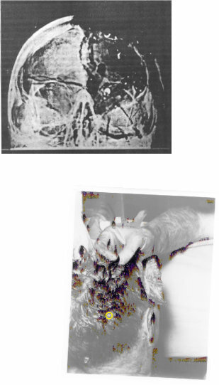
Coming back to the bright object which does not appear to have any bottom in the brightness-coded autopsy picture, the figure below depicts the location of the bright spot on the overlay of original photograph and the colour-coded "bottom" (dark tones), and the flipped X-ray image of President's head. While the head picture and the X-ray do not have identical orientations, it is still possible to at least evaluate the approximate distance of the bright 6.5 mm spot in the X-ray (which we know is only about 2.5 wide, as per Dr Mantik's observation) from the midline and to compare it with the bright looking spot not having a dark bottom in the autopsy picture.
-
From: JFK Absolute Proof, by Robert Groden, page 151.
"There was a massive wound at the back of his head. I recall the injury being right along this area. There was a big hole there. There was a large hole in this area".

-
I have no doubts about the veracity of reports of Parkland doctors and a nurse (Audrey Bell) on the occipital or occipital-parietal location of the main head wound. They would not mistake the occipital-parietal location for a frontal-temporal flap that none of them had seen. For them to make such a mistake they were too much of experts. Parkland doctor's reports are not comparable in strength to e.g., witness testimonies on the number of shot or their directions, which obviously are subject to perceptual errors. Any surgeon having the opportunity to view JFK's head from a short distance (e.g., 2 feet) and for a reasonable period of time (minutes) would be able to describe the location of the head wound(s). This is the reason for the highly convergent reports across the group of surgeons attending the JFK in trauma room 1.
However, several people in Bethesda alo reported to have seen occipital-parietal head wound, e.g., the chief radiologist at the Bethesda Naval Hospital, Dr Ebersole.
-
1 hour ago, Sandy Larsen said:
Problem is, Kennedy had turned his head to the right.
Sandy: are you sure about the right-side turn? I thought there was a slight forward slump of the head and then violently to the back and left.
-
Steven:
did Zapruder's interview happen before or after he had a chance to view his film?
-
I understand that Zapruder's gesture to indicate the location of the head wound and the direction of the spatter was not accurate. It cannot compete with a large number of reports by the Parkland surgeons who had time to observe the head wound from a close distance and for a long periods of minutes. Further, the missing piece of bone known as Harper fragment could not originate from the frontal bone; it was most likely a piece of occipital bone although I would place it slightly differently in occipital bone compared to Dr Mantik's reconstruction. The occipital location of Harper fragment would match the reports of Parkland staff accurately. Notably, that missing fragment in occipital bone is nowhere to be seen in the autopsy photographs.
-
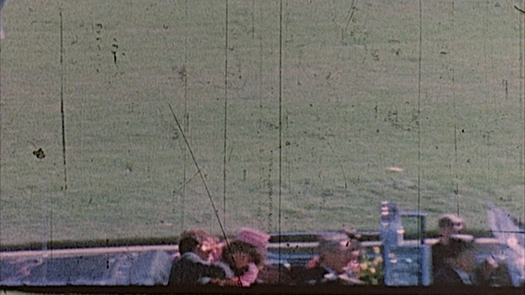
This frame 313 is from a 6k copy of a 3rd generation (32 mm) copy of Zapruder film, posted on midnightwriternews.com. The colours are in logarithmic scale. The black blob in the back of the head appears as a crude alteration aimed to cover for the large wound in the right occipital-parietal area. Difficult to dispute this alteration in my view.


Prayer Man
in JFK Assassination Debate
Posted
The point of my post was that Hosty's statement written not on any sheet of paper but on a sheet he admitted to have taken in Fritz's office was authentic. Whether Hosty scribbled his notes on this sheet of paper while in Fritz's office or transcribed them moments after the interrogation session on this sheet of paper is tangential to the question of whether Lee Oswald told them during the 3.15 PM session that he went out to watch the P. parade.
The other point I developed in my post was that it was possible for Lee Oswald to respond to the noise of the crowd he heard while sitting in Domino room and move toward the front of the building and into the doorway in time to be filmed by Dave Wiegman. That would be the scenario that Agent Hosty described in his hand-written notes bearing the DPD affiliation.